Spigelian hernia
Spigelian hernia accounts for about 2% of all hernias and, therefore, is relatively infrequent. In 1645, Adriaan van der Spiegel, a Flemish anatomist, first described a defect in the semilunar line (linea Spigeli).
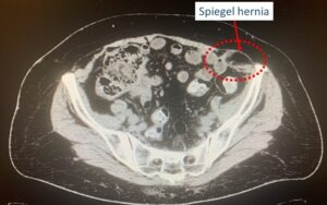
CT scan image of a Spiegel hernia protrusion
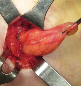
Speigel hernia protrusion after dissection of adhesion bridles.
It can be congenital or acquired, and a wide range of pathologies may act as predisposing factors. Although multiple conditions predispose to developing Spigelian hernias, recent reports have found that at least 50% of all patients with Spigelian hernias had previously undergone abdominal surgery including both open and laparoscopic operations.
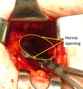
The Spigelian protrusion with its content just pulled back to the peritoneal cavity with a forceps prior of the hernia defect closure
Surgical approaches to Spigelian hernias have evolved over the years, with options ranging from traditional open hernia repair using primary sutures or mesh to more recent laparoscopic techniques, which have gained popularity in the past two decades. In modern times, prosthetic repair is universally regarded as the gold standard for Spigelian hernias, whether performed via open or laparoscopic methods.
Prosthetic repair requires implant fixation to prevent migration.
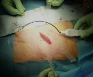
The tentacle shaped mesh Octomesh XS positioned over the incision prior of deployment in a Spiegel hernia repair to demonstrate the wide overlap upon the hernia defect
However, anchoring the mesh within the myotendinous structure of the abdominal wall carries the risk of post-operative complications like bleeding or hematomas. Moreover, the use of sutures or tacks to secure the implants can cause tissue damage, leading to mesh detachment and potential migration, which increases the likelihood of hernia recurrence.
In prosthetic hernia repair, adequate mesh overlap over the defect is crucial. Over time, synthetic materials tend to shrink, potentially exposing the defect and increasing the risk of recurrence. Ensuring substantial defect coverage is especially vital in the case of Spigelian hernia repair.
Given these considerations, Prof. Amato has developed an innovative technique for managing Spigelian hernias. This technique involves using a newly designed tentacle mesh called Octomesh XS, which is intended for fixation free preperitoneal sublay placement using an open approach.
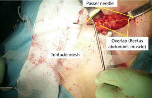
One tentacle of the Octomesh XS is inserted into the eye of a proprietary needle passer to be delivered across the muscle to the preperitoneal space. Of note the broad defect overlap granted by this procedural approach
The Octomesh XS implant features a central oval body with eight bands, or “tentacles,” incorporated along its edge. These tentacle straps are passed through the musculature and fascia to the subcutaneous layer via a specific needle passer in the preperitoneal space.
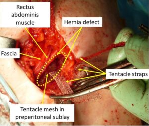
All tentacle straps have been delivered across the abdominal muscles from the suprafascial aspect to the preperitoneal space.
This innovative approach allows for the rapid and fixation-free deployment of the implant, ensuring extensive overlap over the hernia defect.
The friction created by the tentacle straps traversing the abdominal musculature secures the mesh in place, eliminating the need for suture stitches or tack fixation.
The force exerted by these straps, tunneled through the tissues, is strong enough to prevent mesh dislocation.
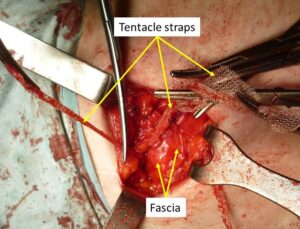
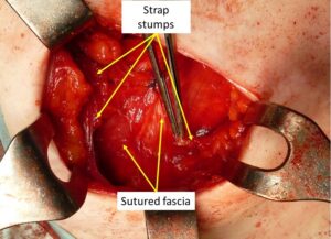
After fascia closure all straps are cut short one by one
This principle’s effectiveness has already been confirmed through experimental testing on a large animal model, (1)
The tentacle mesh is currently being used for surgical repair of incisional, umbilical, and ventral hernias.